Congratulations, Francois, on yet another medical advance. Can you please give us an overview of The Studio?
The Studio offers customized medical and scientific illustrations to anyone who wants them – faculty members of Brown University Health and the Alpert Medical School at Brown University, clinical faculty at other local and regional institutions, researchers, medical technology companies, and anybody who is in need of visual
It is a collaboration with the Brown Imaging Core (BIC), in the Department of Radiology at Rhode Island Hospital. The BIC already provides state-of-the-art radiology-related services for clinicians and researchers (like imaging and video rendering, post-processing manipulation, AI and radiomics). The proximity of radiology capabilities is unique, in that it creates a tight collaboration between MRI and CT scan specialists, computer experts and medical illustrators.
What was the inspiration?
I have been interested in medical illustration for a long time – first, as a user (medical learners and clinicians, and surgeons in particular, rely a lot on visual aids, from anatomy plates to surgical atlases and video libraries. A dilettante artist myself, I have been teaching medical illustration to students and faculty for more than a decade now. With a few allies at the Rhode Island School of Design (RISD), we had explored the idea of developing a graduate program in medical illustration in Providence (the close proximity of Brown and RISD would make this a natural), but that is a long game. In the meantime, we realized that our own faculty and researchers did not have good resources available when they needed to illustrate their grants, their manuscripts or their conference presentations. It turns out that there was an immediate need for these services, and a chance discussion with Dr. Mahesh Jayaraman, Chair of the Department of Radiology, solidified that idea. His department’s BIC already provided imaging-related services to the medical community, and scientific illustration was a perfect addition to their portfolio.
When did work begin on it?
The idea has been germinating for almost two years. Emily Slapin, a certified medical illustrator, had been instrumental in the early years of my medical illustration course. After a BFA at RISD and an MFA at the New York Academy of Art, she attended the oldest, and most famous graduate program in medical illustration in North America, the Department of Art as Applied to Medicine at Johns Hopkins School of Medicine. When she moved back to Providence, she and I immediately thought about collaborating on what would become The Studio. In addition to Emily and Dr. Jayaraman, we were extremely lucky to come in contact with two other collaborators in town, both professional medical illustrators: Lauren Lake and Raisa Rodriguez Maldonado. The final push came in the form of funding from the Research Department at Brown University Health: as an academic researcher himself, Dr. Bharat Ramratnam (Chief science officer at Brown University Health) understood the need for the clinical and research community to have access to high-quality scientific visualization (“SciViz”) tools. This grant allowed us to recruit an administrator for the Studio, to coordinate the interactions between clients and illustrators.
The Studio team includes you and eight others. We will link to them, but can you describe their collective expertise?
In addition to Emily Slapin, Lauren Lake and Raisa Rodriguez Maldonado, who have complimentary interests in medical illustration and animation (ranging from anatomic images to cellular and molecular processes), we have one of the country’s most expert diagnostic imaging computer specialists in Scott Collins. Cross-sectional diagnostic images, like CT scan and MRI, can be further manipulated to create the most amazing, detailed 3D representations of the human body and its various organs and structures. While these semi-automated reconstructions are masterworks of beauty and accuracy in their own right, the collaboration with scientific illustration makes the final result much greater than the sum of its parts: these computer-generated images can form the basis for even more advanced, didactic and accurate illustrations. As an example, we have already been able to provide invaluable information on several sets of conjoined twins early in pregnancy to two medical teams in Minnesota and Wisconsin – information that would not have been possible with ultrasound or even fetal MRI alone. The Studio’s administrator is Lynda Andrade, MBA. Wendy Smith, BS, RT and Grayson Baird, PhD form the link with the research branch of the Department of Radiology.
You state on the website that “a picture is often worth a thousand words, whether it is to write a grant, publish a paper, explain a surgical procedure or hand out patient education. It is all part of visual communication, a way to be better understood. Clear visuals can improve accuracy, engagement and comprehension. They can reduce misunderstandings, speed up decision-making and enhance the learning experience for diverse audiences.” Can you please elaborate?
Most of us are visual creatures – showing is often better than telling when it comes to learning and retaining knowledge. That is true for students (of all ages), and it is also true when we communicate with patients. Medical information can be very dense and filled with long words and complicated concepts. It is often easier to get a point across with an illustration, and patients often keep even the simplest drawing on a napkin as a memento and a “crutch” to remind them of what the doctor said. Illustrations can also cut across languages and even cultural divides. In fact, even the most literary among us probably balk at reading long preoperative or hospital discharge instructions (the social media term TL;DR, or “too long; didn’t read” comes to mind) and prefer short paragraphs with illustrations.
You also write on the site that “The Studio offers resources for clinicians, researchers, educators, developers and entrepreneurs – and anyone who needs a clear picture (or two) to complement, or even replace wordy explanations.” Again, please expand.
Simple drawings or scribbles on a whiteboard can be useful during doctor-patient interactions or when teaching students, but there is a place for elaborate, detailed illustrations in medicine and science. Teaching anatomy to a medical student or learning how to perform an operation requires detailed visuals that are scientifically accurate and educational. A photograph reflects reality, but is not necessarily didactic: the orientation, lighting or focus can be wrong, there may be distracting details or blemishes, and large objects (or organs) may dwarf smaller details. By contrast, a (medical) illustrator can choose the point of view that will best highlight the important aspects of the instruction, mute unimportant details and avoid showing the distracting imperfections that are inherent to all but the most posed studio photographs. An illustration can also show deeper structures by transparency and telescope time and space to show multiple actions, or multiple levels of magnification, in the same image. Creating the perfect medical illustration requires not only artistic talent, but an understanding of the scientific subject, which is further developed through the interaction between the illustrator and the client. To quote the Association of Medical Illustrators, it is not just about beautiful pictures – it is visual problem solving.
And you write that “in addition to static illustrations, we also offer multiple other formats, from interactive and 3D models to animations.” Why are these formats also important?
We live in a 4-dimensional world. Videos are everywhere (anybody can watch a YouTube video of a live operation and learn how to be a surgeon – or so it seems). When used judiciously, animations can be more effective than successive static illustrations, for example to show how micro-organisms attack a human cell or how molecules interact. Computing power is so vast today that realistic, 3D images can be easily created – and artificial intelligence is, of course, ubiquitous. On that note, people often ask why an A.I.-generated image cannot be more useful, and whether it signifies the end of human illustrators. There is absolutely nothing wrong with using these tools to create beautiful medical illustrations – but we should not trade beauty and complexity for accuracy. As of now, A.I. is still fraught with errors and prone to ‘hallucinations.’
View some videos:
— Women’s Wellness and Healthy Aging. Click here.
— NEMO – Neonatal illness. Click here.
— How Does Smell Work? Click here.
You will be offering services to others (for a fee). What schools, centers and others do you expect to be interested?
The Studio’s primary clients are clinicians and researchers, but effective scientific illustrations can also be useful for a tech company promoting its newest device or drug, or a start-up trying to obtain angel funds. Hospitals may need them to develop user-friendly information pamphlets and posters, undergraduate and medical students use plenty of visual didactic material, and even injury-related court cases are often enhanced with vivid medical illustrations. And while we aim to serve our local and regional communities, there is no reason why The Studio couldn’t have a global reach.
Finally, medical illustrations have a long — indeed, ancient — history. How have they helped advance science and the practice of medicine?
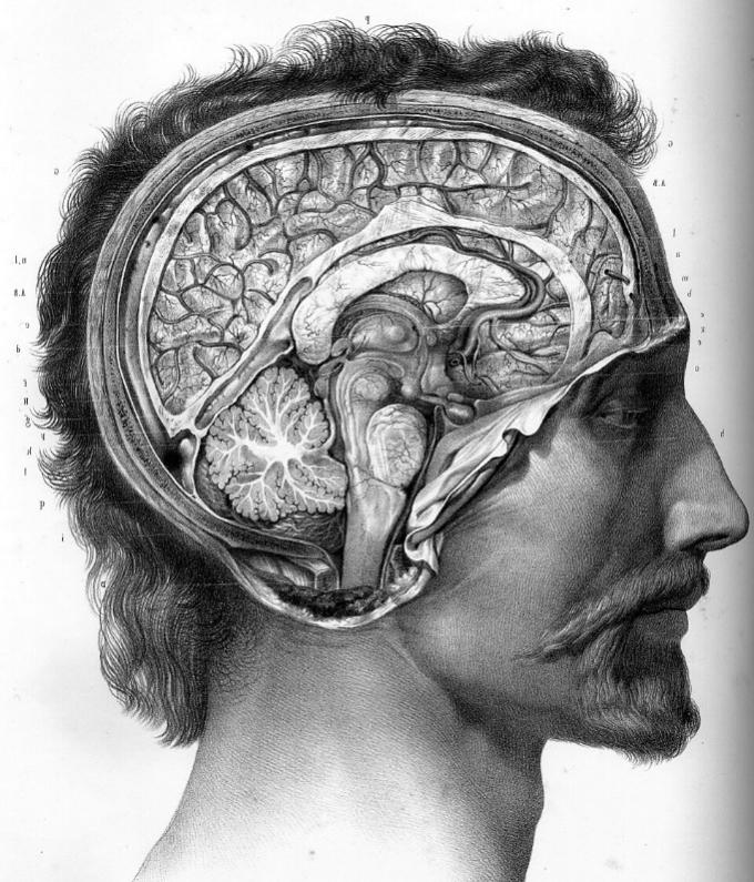
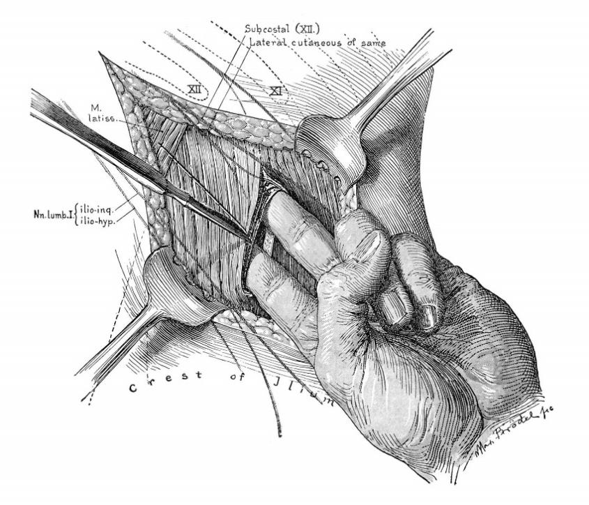
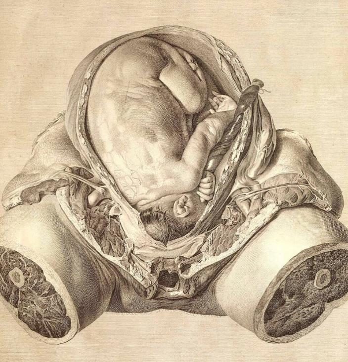
Medical illustration is as ancient as medicine – but modern medicine is only a few centuries old. It started in the Renaissance, with the discovery of human anatomy. The likes of Andreas Vesalius gained widespread popularity thanks to the invention of the printing press – but also because of their use of illustrations (woodcuts, at the time). As anatomy made way for progress in physiology and biology, illustrators were there to document new discoveries. (For example, the description of blood circulation by William Harvey, in the early 1600s, is not much more than three engravings of forearm veins being compressed and released, demonstrating his experiment.) The introduction of general anesthesia and sterile techniques in the late 19th century coincide with a proliferation of surgical atlases, as well as the creation of graduate schools of medical illustration, starting with the Johns Hopkins program. Not only did medical students need to learn about anatomy – now, surgeons needed to share new operative techniques. At the same time, reproduction techniques improved (woodcuts, then engravings, followed by lithography, photography and offset printing – and now, digital publishing). Interestingly, the availability of high-resolution photography and video have not replaced well-planned, didactic medical illustrations. Today, medical illustration also includes that which cannot be seen with the naked eye: think about the ubiquitous 3D illustration of the COVID virus.
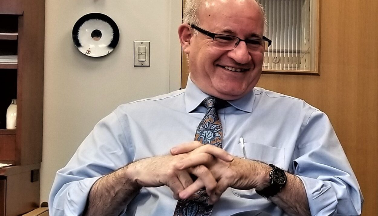
Editor’s note: Among his many other roles, Dr. Luks is Pediatric Surgeon-in-Chief at Hasbro’s Children’s Hospital and course director of Brown University’s PLME 0400, Introduction to Medical Illustration.